Hashimoto's Stages and Diagnosis
Hashimoto's thyroiditis is a chronic autoimmune disorder in which the immune system attacks the thyroid gland, and the 3 diagnostic indicators that confirm it are a positive anti-thyroid peroxidase antibody (anti-TPO) result, a positive anti-thyroglobulin antibody (anti-Tg) result, and a thyroid ultrasound showing a diffusely heterogeneous or coarsely echogenic gland. It is the most common cause of primary hypothyroidism in iodine-sufficient regions, accounting for roughly 80% of all primary hypothyroid cases in the United States, and it affects an estimated 4.6% of adults in the US when antibody testing is included, a rate that climbs to about 14% among women aged 60 and older. A diagnosis of Hashimoto's is made by combining those antibody markers with thyroid function values and, in many cases, a thyroid ultrasound, and the pattern of results shifts as the disease moves through its 3 recognizable clinical stages.
Because antibody positivity and thyroid function can drift independently over years, a patient who tests positive for anti-TPO antibodies while still showing a normal TSH level sits in a distinct category sometimes called Hypothyroidism with Normal TSH, and the management guidance for that category differs from the guidance given once TSH has crossed into the subclinical or overt range. Learning to read those panels yourself, stage by stage, is the core skill this page builds, and it is the starting point for every deeper question about Understand your Hashimoto's trajectory, from first immune attack to a stable replacement regimen.
What Hashimoto's Actually Is and Why It Counts as an Autoimmune Disorder
Hashimoto's thyroiditis is classified as an organ-specific autoimmune disease, and the mechanism is well mapped: T-helper cells, driven by a combination of HLA-DR3 and HLA-DR4 haplotypes and a triggering stimulus such as a viral illness or severe iodine load, direct B cells to produce antibodies against 2 thyroid proteins, thyroid peroxidase (TPO) and thyroglobulin. The antibodies themselves do most of the early damage by triggering antibody-dependent cellular cytotoxicity, in which natural killer cells destroy thyrocytes that have TPO or thyroglobulin on their surface. The end result is a patchy, then progressive, loss of functional thyroid tissue, and the clinical expression of that loss is what a patient experiences as the disease moving from stage to stage.
It is also important to note the distinction that patients often blur: Hashimoto's and hypothyroidism are related but not identical. A person can carry the autoimmune attack for years and remain clinically euthyroid, meaning the thyroid compensates for the tissue loss by upregulating TSH-stimulated output. Hypothyroidism is the functional state; Hashimoto's is the immune mechanism that, in most iodine-sufficient patients, produces it. You can have the autoimmune disorder without yet having the hypothyroid state, and you can develop hypothyroidism from other causes, such as post-surgical thyroidectomy or radioactive iodine ablation for Graves' disease, without any autoimmune thyroiditis at all.
The 3 Clinical Stages of Hashimoto's Thyroiditis
The 3 stages of Hashimoto's thyroiditis describe the progression from first immune infiltration to established hypothyroidism, and each stage carries a distinct antibody and TSH profile that a lab panel will show.
| Stage | Typical Duration | TSH Range | Anti-TPO | Clinical State | Key Finding |
|---|---|---|---|---|---|
| Stage 1: Euthyroid autoimmune thyroiditis | 2 to 10 years, sometimes longer | 0.4 to 4.0 mIU/L (normal) | Positive, often 300 to 1,000+ IU/mL | No symptoms or mild, nonspecific ones | Antibodies are the only abnormality; ultrasound may show heterogeneity |
| Stage 2: Subclinical hypothyroidism | 1 to 5 years | 4.0 to 10.0 mIU/L (mildly elevated) | Positive, often rising | Fatigue, cold sensitivity, mild weight gain of 2 to 5 kg | T4 still within reference range; TSH is the first value to shift |
| Stage 3: Overt primary hypothyroidism | Progressive, often permanent | 10.0 to 100+ mIU/L | Positive, variable | Full hypothyroid symptom spectrum | Free T4 falls below 0.9 ng/dL; levothyroxine replacement begins |
A small subset of patients, roughly 3% to 5% in early series, present with a brief thyrotoxic "hashitoxicosis" phase in which the initial wave of follicular destruction releases stored T4 and T3 into the bloodstream, producing a transient TSH suppression to below 0.1 mIU/L before the gland settles into the hypothyroid pattern. That spike is a one-time event, not a stage a patient manages, and it resolves in 4 to 8 weeks as the stored hormone pool is depleted.
The Full Symptom Spectrum of Hypothyroidism from Hashimoto's
The symptom spectrum of hypothyroidism caused by Hashimoto's thyroiditis spans the nervous, cardiovascular, metabolic, integumentary and reproductive systems, and the pattern below is drawn from the 12 most commonly reported signs in primary-care and endocrinology cohorts.
- Fatigue and reduced exercise tolerance, present in 70% to 90% of overt cases
- Cold intolerance, often noticed first in the hands and feet during autumn months
- Weight gain of 2 to 5 kg over 6 to 12 months, driven by slowed metabolic rate rather than overeating
- Dry, coarse skin and non-pitting edema (myxedema) in the face, eyelids and shins
- Constipation, reflecting reduced gastrointestinal motility
- Menstrual irregularity, including menorrhagia or oligomenorrhea, in reproductive-age women
- Elevated serum LDL cholesterol by 10 to 30 mg/dL, a direct consequence of reduced hepatic LDL-receptor activity
- Hoarse voice, from myxedematous infiltration of the vocal cords
- Cognitive slowing, poor short-term recall, and a subjective sense of "brain fog"
- Bradycardia, with resting heart rate dropping below 60 beats per minute in severe cases
- Depressed mood, ranging from low-grade dysphoria to major depressive episode
- Thyroid goiter in 10% to 30% of patients, a diffuse, rubbery enlargement palpable in the anterior neck
Not every patient presents with all 12 signs. In Stage 2, a patient may notice only 2 or 3 of them, such as fatigue and cold intolerance, while the remaining 10 stay subclinical. The symptom load builds as free T4 declines, and a clinician uses that pattern, not a single isolated sign, to correlate the subjective report with the lab panel.
How Hashimoto's Hypothyroidism Is Tested and Diagnosed
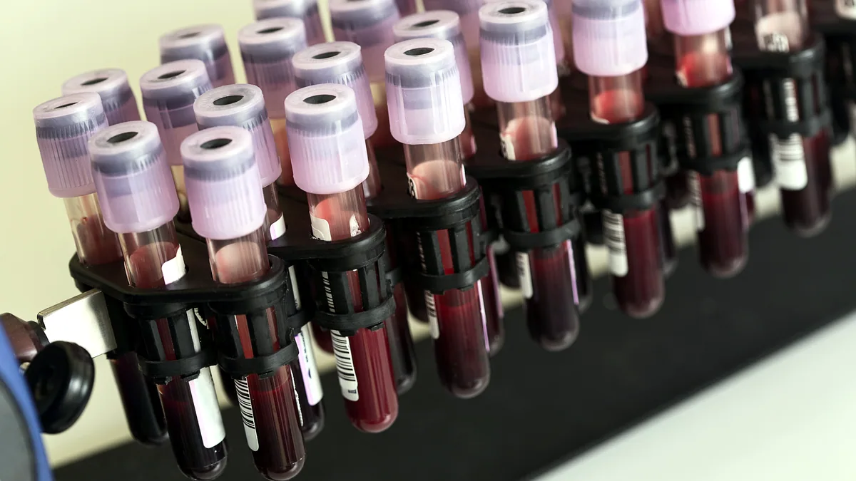
To test for Hashimoto's hypothyroidism, a clinician orders a 3-panel blood draw: thyroid-stimulating hormone (TSH), free T4 (FT4), and anti-TPO antibodies, and in some protocols adds free T3 and anti-thyroglobulin antibodies for a more complete picture.
Reading the Panel
The TSH assay is the most sensitive first-line test because the pituitary amplifies small changes in circulating T4; a drop in free T4 of even 5% can double the TSH. The reference range for TSH on most modern 3rd-generation assays is 0.4 to 4.0 mIU/L, though some endocrinologists use a tighter 0.5 to 2.5 mIU/L range for monitoring patients on replacement. Free T4 typically falls in the 0.8 to 1.8 ng/dL window on immunoassay platforms, and free T3 sits around 2.0 to 4.4 pg/mL. Anti-TPO antibodies above 34 IU/mL on a Roche or 9.0 IU/mL on an Abbott assay are considered positive, and a titer above 1,000 IU/mL is strongly associated with progression to overt hypothyroidism within 5 years.
Imaging and Biopsy
A thyroid ultrasound is the standard imaging step. In Hashimoto's, the sonographer reports a diffusely hypoechoic gland with a coarse or heterogeneous echotexture, sometimes with small echogenic septa, and the gland may measure 15 to 25 mL (normal adult female volume is about 13 to 18 mL). A fine-needle aspiration biopsy is reserved for a discrete nodule that appears suspicious on ultrasound, typically one larger than 1.5 cm with microcalcifications or irregular margins; biopsy is not required to confirm the autoimmune diagnosis itself.
The diagnostic sequence is straightforward: if TSH is elevated and anti-TPO is positive, the diagnosis is confirmed without imaging. If TSH is normal but anti-TPO is positive, the patient is in Stage 1 and is monitored with repeat panels every 12 months. If TSH is elevated but antibodies are negative, the clinician looks for non-autoimmune causes before labeling the picture as Hashimoto's.
Does Hashimoto's Always Get Worse, and How the Numbers Move Over Time
Hashimoto's thyroiditis is a progressive autoimmune process, and the gland's functional reserve declines on average by 10% to 15% per year once the autoimmune attack is established, which is why the trajectory from Stage 1 to Stage 3 is expected rather than exceptional. The key word in that statement is "on average": roughly 25% to 30% of patients with positive antibodies and a normal TSH never progress to overt hypothyroidism during a 10-year follow-up window, while another 5% to 10% progress within 2 years. The rate of progression is influenced by the initial anti-TPO titer, the degree of glandular heterogeneity on ultrasound, and the presence of a concurrent goiter.
Tracking your own numbers over time is where the patient's understanding shifts from passive to active. The minimum monitoring cadence is:
- Stage 1 (euthyroid, antibody positive): TSH, FT4, and anti-TPO every 12 months, with an ultrasound every 24 months
- Stage 2 (subclinical): TSH and FT4 every 6 months for the first 2 years, then every 12 months if stable
- Stage 3 (on levothyroxine): TSH 6 to 8 weeks after any dose change, then every 6 to 12 months at a stable dose; FT4 checked annually
When you read your own panel, watch 3 things: the TSH trend across 2 or 3 draws (a steady climb of 0.5 to 1.0 mIU/L per year is the signal that the gland is losing reserve), the free T4 trajectory (a drift below 1.0 ng/dL in a previously euthyroid patient marks the transition to subclinical hypothyroidism), and the anti-TPO titer (a sustained titer above 1,000 IU/mL, as reported in cohort studies by Vanderpump and colleagues at the University of Oxford and by Biondi and colleagues at the University of Catania, correlates with a faster slide into Stage 3). A single out-of-range value is not a diagnosis; the pattern across 2 or more draws is what carries clinical weight.
Hashimoto's Encephalopathy and the Rare Severe End of the Spectrum
Hashimoto's encephalopathy, also called steroid-responsive encephalopathy associated with autoimmune thyroiditis (SREAT), is a rare neurological complication in which the same anti-TPO antibodies that attack the thyroid cross into the central nervous system and provoke an encephalopathic syndrome. It occurs in fewer than 1 in 100,000 person-years in the general population, and the clinical picture ranges from a subacute confusional state with myoclonic jerks to a seizure disorder or a psychiatric presentation mimicking bipolar disorder or schizophrenia. What distinguishes it from other encephalopathies is the response to glucocorticoids: a high-dose prednisone or methylprednisolone taper typically reverses the neurological symptoms within 7 to 21 days, which is why the condition is labeled "steroid-responsive." The thyroid function at the time of presentation is variable; some patients are euthyroid, some are hypothyroid, and a small number are mildly thyrotoxic, so the diagnosis rests on the antibody titer, the exclusion of other encephalitis causes, and the steroid response rather than on the TSH value alone.
The encephalopathic presentation is the most extreme expression of the autoimmune process that defines Hashimoto's thyroiditis, and it is the reason clinicians take a positive anti-TPO titer seriously even when the thyroid panel looks normal. For the vast majority of patients, however, the disease follows the 3-stage pattern outlined above, the symptom spectrum stays manageable with levothyroxine, and the annual panel tells you everything you need to know about where your gland stands and what the next 12 months are likely to look like.